Introduction
Cancer research has been a dynamic and ever-evolving field, with scientific advancements continually reshaping our understanding of tumorigenesis and offering new avenues for therapeutic intervention. One of the most intriguing and promising developments in recent decades has been the utilization of small fish models, such as zebrafish (Danio rerio), for live-imaging analyses. As detailed in an article published in the “Yakugaku Zasshi: Journal of the Pharmaceutical Society of Japan,” these models have significantly propelled our knowledge of the mechanisms regulating primary tumorigenesis (Haraoka et al., 2019). In this news article, we delve into the insights provided by zebrafish models and how they are revolutionizing cancer research and drug discovery.
The Value of Zebrafish in Cancer Research
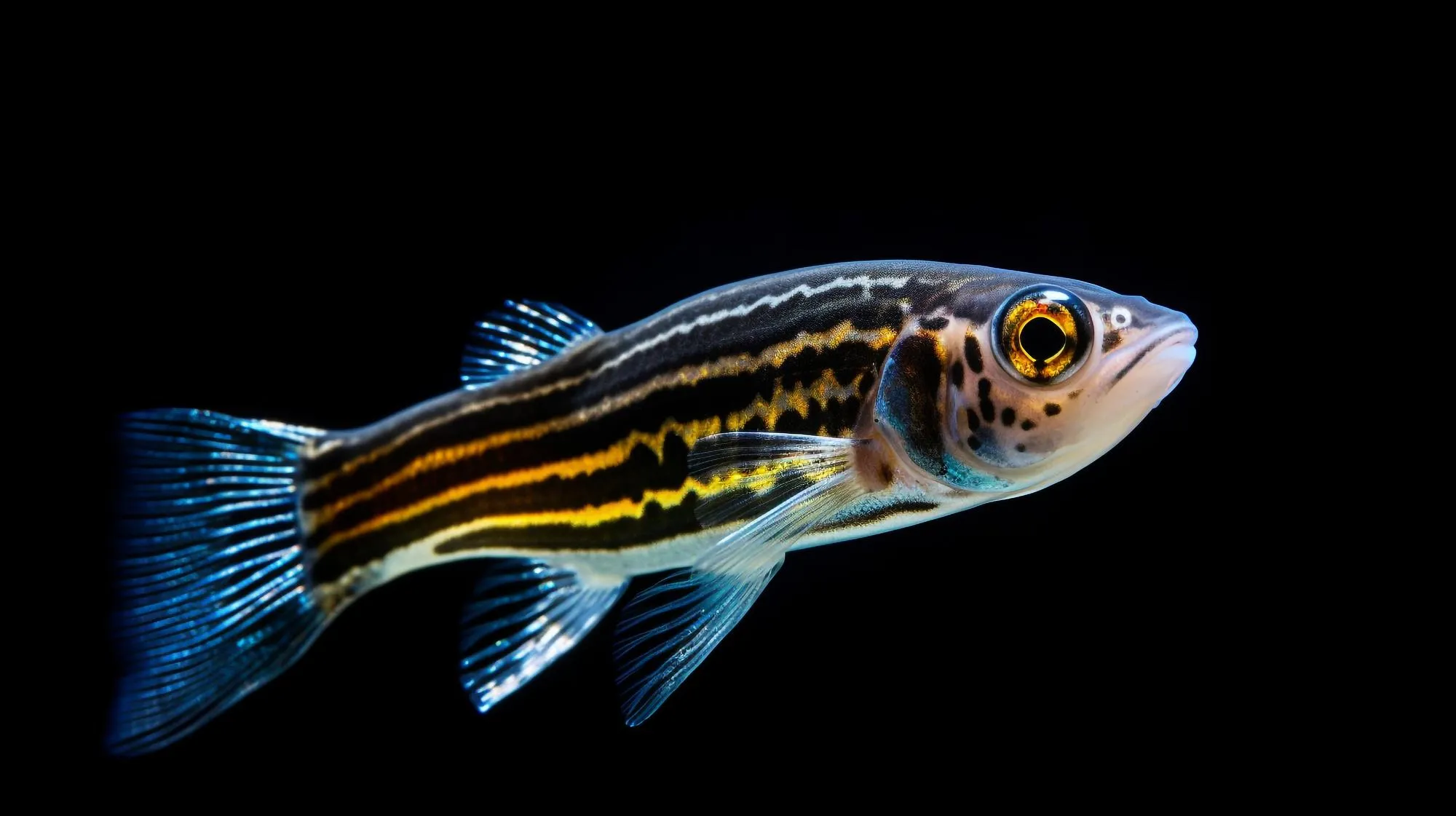
Since the 1980s, the zebrafish has been a valuable model system for investigating various developmental processes. This small tropical fish possesses three key characteristics that make it particularly advantageous for scientific study: its embryos grow outside the mother, providing easier observation and manipulation; the embryos are transparent during the early stages of development, allowing for direct imaging of internal processes; and zebrafish have organs and cellular pathways that are remarkably similar to those in humans (Haraoka et al., 2019).
Beyond their use in developmental biology, zebrafish have emerged as a powerful tool for studying human diseases, especially cancer. What gives zebrafish a unique edge over other model organisms, such as flies, mice, and rats, is their low-cost breeding, suitability for high-throughput studies, high efficiency of transgenesis, and the ease with which researchers can observe cancer progression, invasion, and metastasis in vivo (Haraoka et al., 2019).
Harnessing Zebrafish for Cancer Mechanism Insights
The profound transparency of zebrafish at the embryonic stage coupled with advanced imaging technologies allows researchers to visualize tumor development and metastasis in living organisms—something that was previously unattainable with traditional models. This capability has driven significant progress in understanding the dynamics of cancer cells within their native environment, leading to the discovery of novel mechanisms underlying tumorigenesis and metastasis (Haraoka et al., 2019).
For example, live-imaging studies using zebrafish have revealed intricate interactions between tumor cells and the surrounding microenvironment, which plays a crucial role in cancer progression. The interactions between cancer cells and host tissues, such as blood vessels and immune cells, can be observed in real-time, providing invaluable insights into the processes of tumor angiogenesis, immune evasion, and the pre-metastatic niche formation (Haraoka et al., 2019).
Accelerating Drug Discovery with Zebrafish Screens
The potential of zebrafish extends to the domain of drug discovery. The species’ small size and compatibility with high-throughput screening techniques have enabled researchers to rapidly test thousands of compounds, shortening the timeline for identifying promising anticancer drugs. Zebrafish larvae can be immersed in small wells containing different chemical compounds, and their responses are observed using high-content imaging systems. This approach has already identified numerous potential therapeutic agents that may inhibit tumor growth, angiogenesis, and metastasis (Haraoka et al., 2019).
Zebrafish models have also proven useful in unraveling the genetic basis of cancer. With ease of genetic manipulation, scientists can introduce human cancer-related genes into zebrafish and observe the consequent effects on tumorigenesis. This valuable inter-species gene function analysis can shed light on the pathogenicity of variants detected in human patients, providing additional layers of data for precision medicine approaches (Haraoka et al., 2019).
Challenges and Future Directions
While the advantages of zebrafish models in cancer research and drug discovery are clear, challenges remain. Not all human cancers can be modeled accurately in zebrafish, and differences in immune system complexity and tumor microenvironment between zebrafish and humans may lead to divergent drug responses. Nonetheless, as technologies continue to advance, these models will undoubtedly become increasingly sophisticated, allowing for more precise recapitulation of human diseases.
Moreover, current research efforts focus on enhancing the utility of zebrafish for pan-cancer studies, encompassing various cancer types and subtypes. Through CRISPR-Cas9 and other genetic engineering techniques, zebrafish are being equipped with complex genetic alterations that mirror human mutations, thus creating more predictive models of cancer biology and response to therapy.
Conclusion
The review article by Haraoka et al. (2019) in “Yakugaku Zasshi” underscores the substantial contributions of zebrafish to cancer research. As we continue to explore and exploit the unique features of this small fish model, scientists are optimistic about its role in accelerating the discovery of novel cancer mechanisms and treatments. The zebrafish’s journey from a pet store novelty to a pillar of cancer research is a testament to the transformative power of innovative scientific tools in understanding and combating this multifaceted disease.
