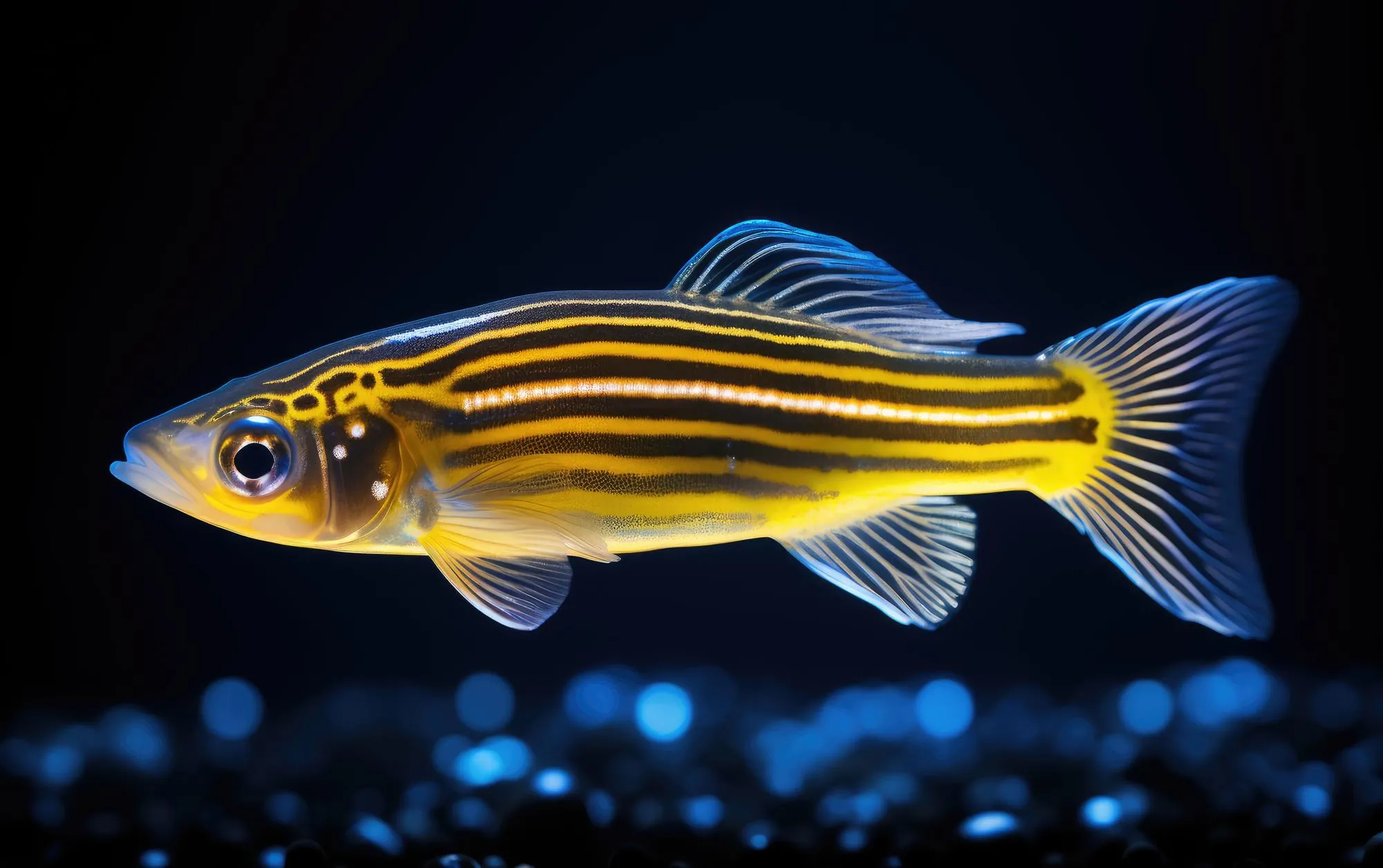
A team of researchers from Rice University have provided a groundbreaking new insight into the development of the enteric nervous system (ENS), publishing their findings in the journal Scientific Reports. The study, “Immunohistochemical and ultrastructural analysis of the maturing larval zebrafish enteric nervous system reveals the formation of a neuropil pattern” (DOI: 10.1038/s41598-019-43497-9), sheds light on the intricate development of ENS ganglia and the emerging neuropil patterns that form crucial connections throughout the gut.
Abstract of the Evolving Enteric Network
The gastrointestinal (GI) tract is an organ system that runs the length of our body, with its own series of interconnected ganglia called the ENS. These ganglia facilitate critical reflex control of various gut operations, such as peristalsis, water balance, hormone secretions, and intestinal barrier functions. For years, scientists have questioned the developmental mechanisms that guide the organization of the neural networks within the ENS. In this study, the authors utilized the zebrafish model, a valuable organism in neurodevelopmental research, to demystify the intricate formation of the myenteric plexus, one of the primary plexuses in the ENS.
Keywords
1. Enteric Nervous System Development
2. Zebrafish Neural Research
3. Neuropil Pattern Formation
4. Myenteric Plexus Architecture
5. Neuroglia Interactions
The Study in Detail
Phillip A. Baker, Matthew D. Meyer, Ashley Tsang, and Rosa A. Uribe of Rice University’s Biosciences Department conducted a meticulous investigation on the developing zebrafish ENS. Their study spanned immunohistochemical evaluations and advanced electron microscopy to observe the architectural intricacies of the maturing myenteric plexus over time.
In the early stages of development, researchers noticed that tight layers of axons and elongated processes of glial cells were encapsulating the gut tube’s circumference. As the larval stages advanced, the complexity within the myenteric plexus increased, with axon layers juxtaposing concentric layers of glial cells.
Ultrastructural observations further revealed that glial cells contained filaments and formed extensive contacts with one another in lengthy projections. Significantly, indicators of vesicular axon profiles were abundant throughout the larval plexus neuropil, suggesting active development and synaptic potential in these regions.
This discovery aligns with parallel studies in the field, including works by researchers like Le Douarin and Teillet (Development, 1973), where migration patterns of neural crest cells to the digestive tract wall in avian embryos were first described. It is now understood that neural crest cells are progenitors of the enteric ganglia (Epstein et al., Dev. Dyn., 1994), and these cells are pivotal for ENS formation.
Implications of the Study
Understanding the ENS architecture is vital because of its role in various gut-related dysfunctions and diseases, including inflammatory bowel disease and Hirschsprung disease. The research undertaken by Baker et al. opens the door for functional studies to better understand how this intricate network forms and operates, potentially leading to improved treatment for GI conditions.
The realization that the zebrafish ENS and its maturation closely mirror that of humans is also significant, highlighting the zebrafish as an effective model for studying human ENS development and related pathologies (Ganz J., Dev. Dyn., 2018).
Future Directions and Research Potential
The study authors are optimistic about the future of ENS research, emphasizing the potential of the zebrafish model in furthering the understanding of neurogenesis within the gut. According to Uribe et al., follow-up studies could focus on the specific roles of various cell types, including interstitial cells of Cajal and glial cells, and their involvement in ENS pathologies (Uribe RA, et al., Mol. Biol. Cell, 2015).
As hypothesized by Rao and Gershon (Nature Reviews Neuroscience, 2018), disruptions in the typical ENS development could manifest in a myriad of digestive issues. The well-documented architecture and progression of ENS maturity in zebrafish now provide powerful investigative avenues for developmental biologists and neuroscientists.
Broader Relevance and Considerations
This research holds implications beyond academia, potentially impacting pharmaceutical and medical treatments. Understanding the formation and function of the ENS enables drug developers to create more targeted therapies for GI disorders. Moreover, it poses questions about the impact of nutrition, probiotics, and other environmental factors on ENS health and maintenance (Komuro et al., Neuroscience, 1982).
As the study concludes, Baker and the team advocate for a multidisciplinary approach to ENS research, incorporating genetics, developmental biology, and bioengineering. By combining these fields, the potential for innovation and discovery in the treatment of ENS-related conditions is profound.
Clinical Applications and Conclusion
The comprehensive analysis performed by the Rice University team demonstrates the immense complexity of ENS development. Their use of cutting-edge technology to visualize the development of neural networks enables the medical community to address some of the most pressing GI tract diseases.
In future research, clinicians might utilize data on the ENS architecture to manage and treat conditions like GI motility disorders, offering new hope to patients worldwide. The detailed visualization of the ENS at different larval stages provides a template for developmental benchmarks, possibly leading to earlier detection and intervention strategies in diseases affecting the ENS.
References
1. Baker, P. A., Meyer, M. D., Tsang, A., & Uribe, R. A. (2019). Immunohistochemical and ultrastructural analysis of the maturing larval zebrafish enteric nervous system reveals the formation of a neuropil pattern. Scientific reports, 9(1), 6941.
DOI: 10.1038/s41598-019-43497-9
2. Epstein, M. L., Mikawa, T., Brown, A. M. C., McFarlin, D. R. (1994). Mapping the origin of the avian enteric nervous system with a retroviral marker. Dev. Dyn., 201:236–244. doi: 10.1002/aja.1002010307.
3. Ganz, J. (2018). Gut feelings: Studying enteric nervous system development, function, and disease in the zebrafish model system. Dev. Dyn., 247:268–278. doi: 10.1002/dvdy.24597.
4. Rao, M., & Gershon, M. D. (2018). Enteric nervous system development: What could possibly go wrong? Nature Reviews Neuroscience, 19(9):552–565. doi: 10.1038/s41583-018-0041-0.
5. Uribe, R. A., Gu, T., & Bronner, M. E. (2015). Meis3 is required for neural crest invasion of the gut during zebrafish enteric nervous system development. Mol. Biol. Cell., 26(23):3728–40. doi: 10.1091/mbc.E15-02-0112.
Epilogue
As researchers around the globe continue to decipher the complexities of neural development, studies like the one conducted by the team at Rice University serve as pivotal reference points, revolutionizing our understanding of the ENS and ultimately paving the way towards innovative therapies for neurological and gastrointestinal health.
