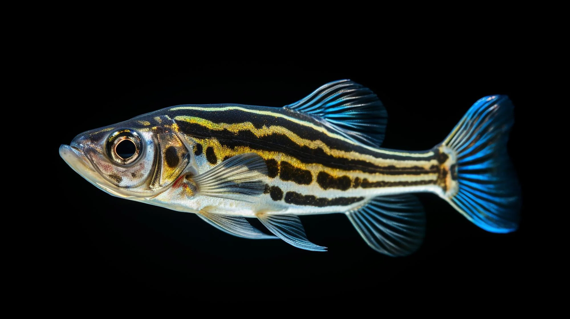
In a groundbreaking study published in the journal Cells & Development, researchers from the Department of Molecular, Cellular, and Developmental Biology at Yale University have leveraged state-of-the-art imaging techniques to explore the dynamic behavior of Fibronectin, a vital extracellular matrix glycoprotein, during the early developmental stages of zebrafish. This study provides a deeper understanding of the complex role that extracellular matrices play in the morphogenesis of living organisms.
The pioneering work, led by Jülich Dörthe D and Scott A Holley, introduces two fluorescently tagged knock-in alleles: Fibronectin 1a-mNeonGreen and Fibronectin 1b-mCherry. These genetically engineered markers allow for real-time in vivo imaging of the fibronectin matrix—a leap forward that overcomes the limitations of traditional fixed-tissue imaging.
Fibronectin is integral to embryonic development, serving as a ligand for several members of the Integrin adhesion receptor family. The Yale University research team’s ambitious approach aims to compare the expression patterns and loss of function phenotypes of two zebrafish fibronectin paralogs, fn1a, and fn1b, providing key insights into their distribution and functionality.
DOI: 10.1016/j.cdev.2024.203900
The live imaging results are striking; Fn1a-mNeonGreen and Fn1b-mCherry co-localize within extracellular matrix (ECM) fibers across the paraxial mesoderm and the myotendinous junction. By using this novel imaging approach, the researchers were able to observe distinct localizations of the two fibronectin forms in the developing zebrafish. Fn1a-mNeonGreen predominated in areas like the branchial arches, heart ventricle, olfactory placode, and otic capsule. Conversely, Fn1b-mCherry was primarily found in the pericardium, proximal convoluted tubule, posterior hindgut, and ventral mesoderm/cardinal vein.
Taking this study a step further, genetic complementation experiments demonstrated that the engineered fluorescent knock-in alleles retain their functionality, thereby validating their use for in vivo studies.
In a series of critical experiments involving maternal zygotic integrin α5 mutants and integrin β1a; β1b double mutants, the paper reveals unique requirements for these integrins in assembling the two fibronectins into ECM fibers within various tissues. This specificity indicates a nuanced interplay between components of the extracellular matrix and their associated integrins during organogenesis.
Rescue experiments, which involved mRNA injection, indicated that Fibronectin 1a and 1b are not perfectly interchangeable, suggesting distinct functional roles for each during development.
A particularly revealing finding of the study was the cross-regulation observed between the two fibronectins. Fn1a proved to be necessary for the normal fibrillogenesis of Fn1b in the presomitic mesoderm, but Fn1b did not exhibit the same dependence on Fn1a for its own normal deposition pattern.
The study’s far-reaching implications underscore the intricate balance and precise regulatory mechanisms governing the embryonic development of vertebrates, including humans.
The research article, which is available ahead of print, confirms there are no competing or financial interests associated with the manuscript, ensuring unbiased and accurate reporting of the findings.
Keywords: Fibronectin, Zebrafish Development, Live Imaging, Extracellular Matrix, Integrin
References
1. Dörthe, J. D., & Holley, S. A. (2024). Live imaging of Fibronectin 1a-mNeonGreen and Fibronectin 1b-mCherry knock-in alleles during early zebrafish development. Cells & Development, 177, 203900. doi:10.1016/j.cdev.2024.203900
2. Astrof, S., & Hynes, R. O. (2009). Fibronectins in vascular morphogenesis. Angiogenesis, 12(2), 165-175. doi:10.1007/s10456-009-9146-2
3. George, E. L., Georges-Labouesse, E. N., Patel-King, R. S., Rayburn, H., & Hynes, R. O. (1993). Defects in mesoderm, neural tube and vascular development in mouse embryos lacking fibronectin. Development, 119(4), 1079-1091.
4. Hynes, R. O. (2009). The extracellular matrix: not just pretty fibrils. Science, 326(5957), 1216-1219. doi:10.1126/science.1176009
5. Trinh, L. A., & Stainier, D. Y. (2004). Fibronectin regulates epithelial organization during myocardial migration in zebrafish. Developmental Cell, 6(3), 371-382. doi:10.1016/s1534-5807(04)00051-2
Keywords
1. Fibronectin Integration Zebrafish
2. Live Cell Imaging ECM
3. Zebrafish Fibronectin Knock-In
4. Zebrafish Developmental Biology
5. Extracellular Matrix Live Tracking
