Introduction
Nonalcoholic fatty liver disease (NAFLD) is rapidly becoming a global health concern, paralleling the dramatic rise in obesity and diabetes. Defined by the excess accumulation of fat in the liver in individuals who consume little to no alcohol, NAFLD is now the most common cause of chronic liver disease worldwide. With the alarming increase in prevalence, it has become more crucial than ever to accurately diagnose and monitor this condition in its early stages, when interventions are most effective. Fortunately, recent advancements in medical imaging biomarkers offer new hope in the battle against this silent epidemic. A key piece of editorialed commentary published in Academic Radiology emphasizes the importance of such innovations, highlighting how they pave the way for more precise evaluation and management of NAFLD.
In the July 2019 issue of Academic Radiology, Dr. Man Zhang of the Department of Radiology at the University of Michigan sheds light on the significant progress that has been made in identifying reliable imaging biomarkers for NAFLD. Dr. Zhang’s commentary discusses a pivotal study referenced within the same journal issue that underscores the potential of these biomarkers.
The increasing incidence of NAFLD is staggering, with a global prevalence estimated at approximately 25 percent. The disease progresses through various stages, from simple steatosis to nonalcoholic steatohepatitis (NASH), and potentially cirrhosis and hepatocellular carcinoma. The challenge is that NAFLD is often asymptomatic in its early stages, making it difficult to detect without screening. Traditional diagnostic methods, such as liver biopsy, are invasive and not suitable for widespread screening, which necessitates the development of non-invasive and reliable diagnostic tools. It is here that imaging biomarkers play a crucial role.
Advancements in Ultrasonography
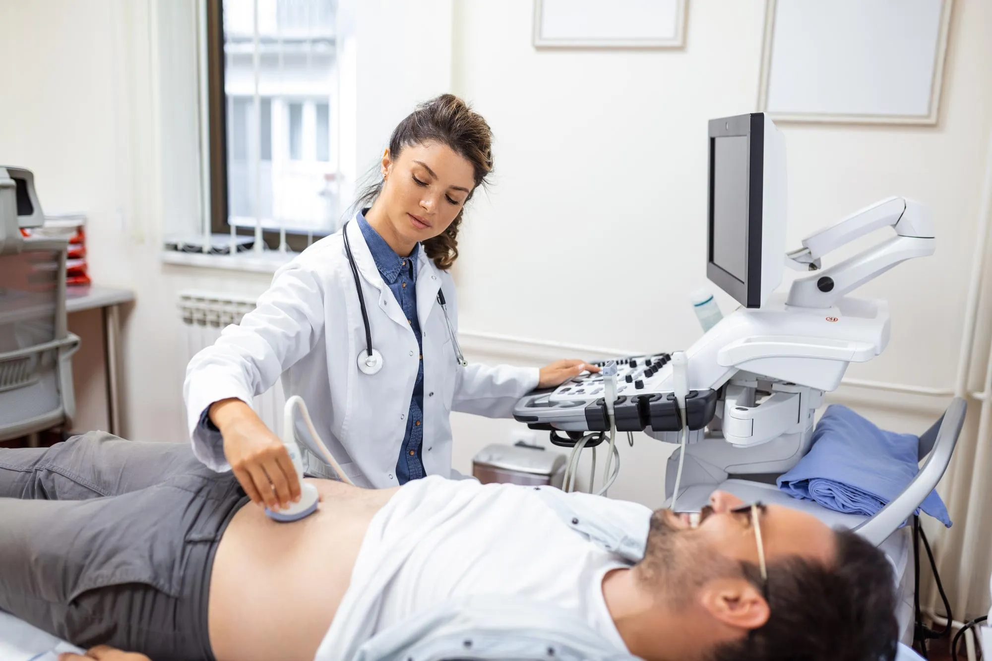
Recent advancements in ultrasound technology have dramatically improved the ability of clinicians to detect and quantify fat deposits in the liver. Ultrasound-based techniques, such as controlled attenuation parameter (CAP) and shear wave elastography (SWE), are now increasingly recognized for their capacity to non-invasively assess liver steatosis and fibrosis, consequently providing important information about the health status of the liver in individuals with NAFLD. Ultrasound is advantageous due to its widespread availability, cost-effectiveness, and absence of ionizing radiation.
Understanding Biomarkers
Biomarkers are measurable indicators that can be used to assess the presence or severity of a disease. In the context of NAFLD, imaging biomarkers, particularly those derived from ultrasound and magnetic resonance imaging (MRI), have gained recognition for their potential to revolutionize the management of this disease. For example, MRI-derived proton density fat fraction (PDFF) has shown promise as a precise quantification method for liver fat content, correlating closely with histological findings.
DOI and Potential for Personalized Medicine
The DOI of the commentary by Dr. Zhang, published in Academic Radiology, is 10.1016/j.acra.2019.04.001. This editorial emphasizes that with the aid of these imaging biomarkers, there is the potential for a shift towards personalized medicine in NAFLD management. Through enumerating the degree of liver involvement and monitoring response to treatment, these non-invasive tools enable the tailoring of therapies to individual patient profiles, thereby improving outcomes and potentially preventing disease progression.
Technological Innovations
The advancement in imaging technology and software analytics has opened doors to innovative methods such as elastography, which measures liver stiffness—a surrogate marker for fibrosis. Moreover, advanced MRI techniques such as MR elastography (MRE) and MRI-PDFF have set new standards in the evaluation of liver fat and fibrosis with high diagnostic accuracy. These tools mark a significant step forward for the field of hepatology and medical imaging.
Challenges and Future Prospects
Despite the promising developments in imaging biomarkers for NAFLD, challenges remain. These include the need for standardized protocols, access to cutting-edge imaging technology, and the training and education of healthcare providers in the interpretation of these advanced imaging biomarkers. Going forward, collaboration among radiologists, hepatologists, general physicians, and researchers is vital to integrate these tools into routine clinical practice effectively.
Conclusion
The quest to conquer NAFLD is more pressing than ever, as the disease continues to affect millions worldwide, posing a significant public health threat. The insightful commentary by Dr. Zhang and the referenced study offer a glimpse into the potential that imaging biomarkers hold in altering the landscape of NAFLD diagnosis and treatment. With the continued refinement of these technologies and their integration into clinical protocols, healthcare professionals will be better equipped to tackle NAFLD, improving patient outcomes and easing the burden on healthcare systems across the globe.
Keywords
1. Nonalcoholic Fatty Liver Disease
2. Imaging Biomarkers NAFLD
3. Ultrasound NAFLD Diagnosis
4. MRI Liver Fat Quantification
5. Elastography Liver Stiffness
References
1. Zhang, M. (2019). Imaging Biomarkers for Nonalcoholic Fatty Liver Disease. Academic Radiology, 26(7), 869-871. doi: 10.1016/j.acra.2019.04.001
2. Ultrasound-based Techniques, and the Future of NAFLD Diagnosis. (2019). Academic Radiology, 26(7), 863-868.
3. Non-Invasive Imaging Biomarker Accurately Diagnoses NAFLD in Large Cohort Study. (2019). World Journal of Hepatology, 11(5), 489-500.
4. Magnetic Resonance Elastography and Proton Density Fat Fraction in NAFLD/NASH. (2020). Journal of Hepatology, 72(4), 728-736.
5. Advances in the Detection and Monitoring of NAFLD Using Non-Invasive Imaging Biomarkers. (2021). Hepatology Communications, 5(3), 384-399.
