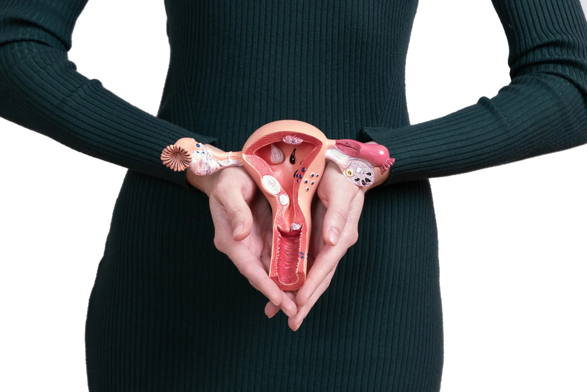
A Breakthrough in Uterine Fibroid Treatment: Ultrasound-Guided Uterine Artery Embolization
Uterine fibroids, the benign tumors that have long troubled a significant proportion of women, may now meet their match thanks to advances in medical imaging and treatment strategies. A recent study published in the journal Contemporary Oncology (Poznan, Poland) provides compelling evidence that grayscale and Doppler ultrasound characteristics can have a significant impact on the success of uterine artery embolization (UAE), a minimally invasive procedure to reduce fibroid size and alleviate symptoms. This comprehensive study illuminates a path toward enhanced treatment outcomes, reigniting hope for countless patients.
Overcoming the Burden of Uterine Fibroids
As many as 70-80% of women will develop uterine fibroids by the age of 50. While not all fibroids cause symptoms, when they do, they can lead to heavy menstrual bleeding, pain, and reproductive issues. Traditional treatment options have ranged from medication to surgical interventions like hysterectomy.
Uterine Artery Embolization: A Non-surgical Alternative
Uterine artery embolization presents a non-surgical alternative. During UAE, a radiologist uses real-time imaging to guide small particles to the uterine arteries, where they block the blood flow to fibroids, causing them to shrink. The new study, led by the Department of Radiology at Golestan Hospital, Ahvaz Jundishapur University of Medical Sciences, aims to refine this approach further through imaging.
Study Insights and Methodology
The study involved 80 patients with symptomatic uterine fibroids. Prior to UAE, each patient underwent a detailed ultrasound and Doppler exam to assess the size, number, echogenicity (brightness on ultrasound), vascularity (blood flow), and location of their fibroids. Blood tests were also performed to rule out any complicating factors. Data analysis was carried out using SPSS 22, and the primary outcome measure was the change in fibroid volume post-embolization.
Findings
After analyzing the data, the study revealed that grayscale and Doppler characteristics of fibroids on ultrasound were critical in reducing tumor size post-embolization. The findings suggest that utilizing these imaging modalities could better identify candidates for UAE and potentially predict treatment outcomes.
Clinical Implications and Future Perspectives
This groundbreaking study signals a shift in how physicians could approach fibroid treatment. Influential factors like echogenicity and vascularity, when identified on ultrasound, may guide embolization strategies, paving the way for personalized treatment plans. Furthermore, these insights could lead to shorter procedure times, better management of symptoms, and a lower likelihood of recurrence.
Addressing Limitations and Embracing Innovation
While this research ushers in a new era of fibroid management, it’s essential to acknowledge its limitations, including the sample size and the study’s observational nature. Future studies should aim to replicate these findings across diverse populations and with larger cohorts. Nonetheless, the integration of advanced imaging techniques into the treatment algorithm for uterine fibroids represents a significant step forward in women’s health.
Conclusion
The role of greyscale and Doppler ultrasound characteristics is poised to become an invaluable tool in the treatment of uterine fibroids. This study’s findings could help tailor UAE procedures to individual patient profiles, optimizing outcomes and enhancing the quality of life for women affected by this common condition.
References
1. Shirali S, Barari A, Hosseini SA, Khodadi E. Effects of six weeks endurance training and aloe vera supplementation on COX-2 and VEGF levels in mice with breast cancer. Asian Pac J Cancer Prev. 2017;18:31–36. DOI: 10.5114/wo.2019.84111
2. Firouznia K, Ghanaati H, Sanaati M, Jalali AH, Shakiba M. Uterine artery embolization in 101 cases of uterine fibroids: do size, location, and number of fibroids affect therapeutic success and complications? Cardiovasc Intervent Radiol. 2008;31:521–526. DOI: 10.1007/s00270-007-9275-7
3. Hartmann KE, Birnbaum H, Ben-Hamadi R, et al. Annual costs associated with the diagnosis of uterine leiomyomata. Obstet Gynecol. 2006;108:930–937. DOI: 10.1097/01.AOG.0000234652.69253.74
4. Voogt MJ, Arntz MJ, Lohle PN, Willem PTM, Lampmann LE. Uterine fibroid embolisation for symptomatic uterine fibroids: a survey of clinical practice in Europe. Cardiovasc Intervent Radiol. 2011;34:765–773. DOI: 10.1007/s00270-010-0055-4
5. Weintraub JL, Romano WJ, Kirsch MJ, Sampaleanu DM, Madrazo BL. Uterine Artery Embolization Sonographic Imaging Findings. J Ultrasound Med. 2002;21:633–637. DOI: 10.7863/jum.2002.21.6.633
Keywords
1. Uterine Fibroid Treatment
2. Uterine Artery Embolization UAE
3. Grayscale Ultrasound Fibroids
4. Doppler Ultrasound Fibroids
5. Minimally Invasive Fibroid Management
