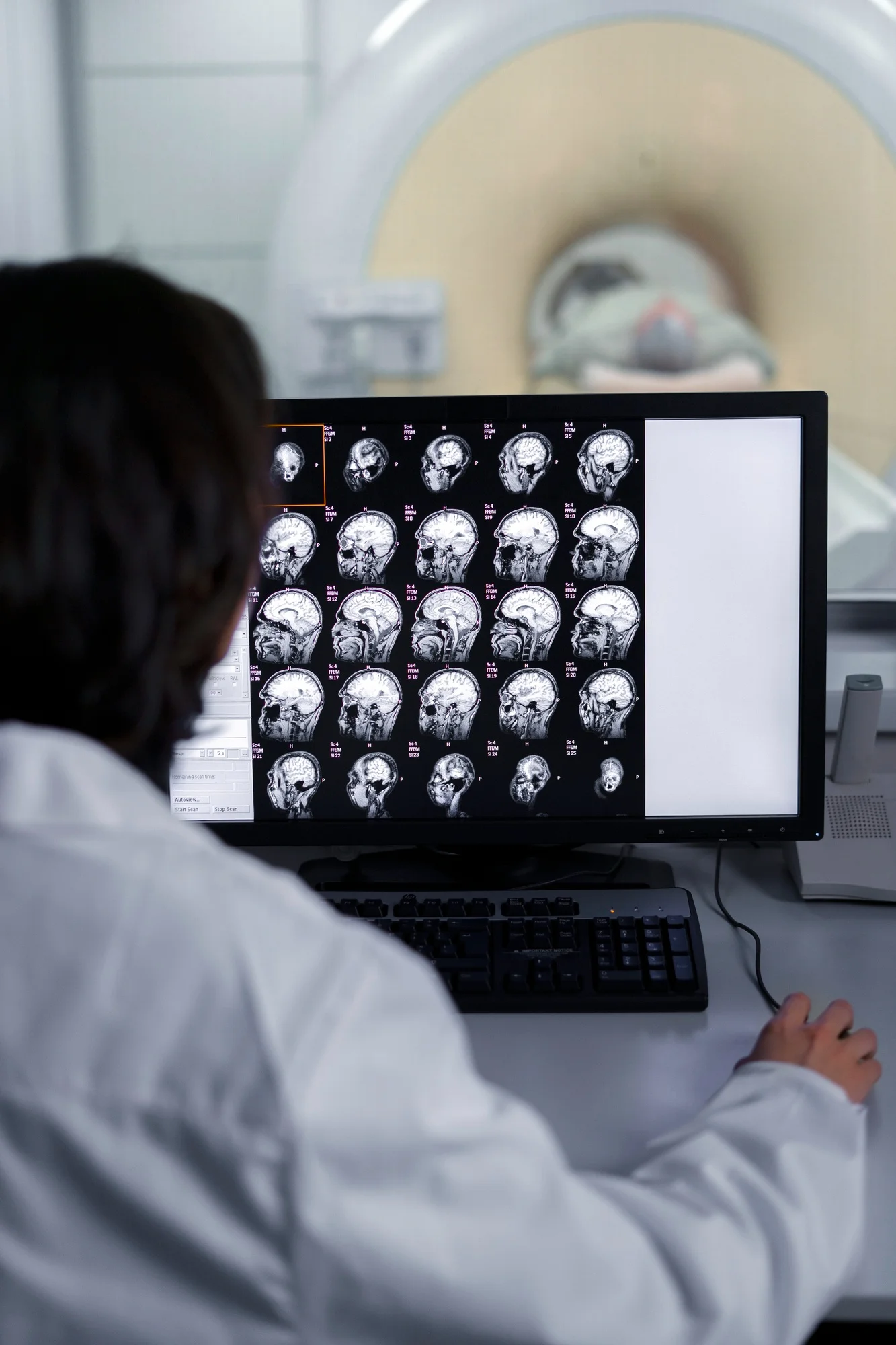
In the continuous effort to improve diagnostic accuracy and patient management in multiple myeloma (MM), a team of researchers from Poland has made a significant contribution. A study led by Czyż Jarosław and his colleagues analyzed the diagnostic accuracy of combined 18F-fluorodeoxyglucose positron emission tomography and computed tomography (18F-FDG PET/CT) in comparison to traditional CT scans in patients with MM. The results, published in Contemporary Oncology (Poznan, Poland), provide critical insights into how imaging techniques can be optimized for the diagnosis and monitoring of MM.
DOI: 10.5114/wo.2019.83342
PMC: PMC6500394
A Novel Insight into Imaging for Myeloma
The clinical trial included a diverse group of MM patients: eight with newly diagnosed active myeloma, nine in very good partial remission (VGPR) or complete remission (CR) after treatment, and 15 with active disease after relapse. The study found that in newly diagnosed cases, conventional CT revealed 64 lesions, while 18F-FDG PET/CT detected 83. The superior sensitivity of 18F-FDG PET/CT was evident for patients in VGPR/CR and those with relapsed disease, indicating that 18F-FDG PET/CT might be the superior imaging modality for MM assessment.
Imaging and MM: A Critical Review
Multiple myeloma, a hematological cancer characterized by the proliferation of malignant plasma cells in the bone marrow, can lead to bone lesions, renal failure, anemia, and hypercalcemia. Accurate detection and evaluation of the disease are vital for effective treatment and management. Traditionally, a combination of laboratory tests, biopsies, and imaging studies are utilized for diagnosis and staging. Imaging studies play an essential role in detecting bone lesions and extramedullary disease, which are significant prognostic factors.
Rajkumar et al. introduced updated criteria for MM diagnosis, emphasizing the relevance of modern imaging techniques such as MRI and PET/CT scans [2]. While CT scans provide detailed images of bony structures, they are not as sensitive as PET/CT for detecting bone marrow involvement or active disease in MM. Furthermore, 18F-FDG PET has demonstrated its potential in evaluating treatment response, guiding biopsy sites, and predicting outcomes in MM [6].
Expert Consensus and Imaging Guidelines
The International Myeloma Working Group (IMWG) champions the integration of modern imaging techniques in MM patient care. The IMWG has recommended the use of whole-body low-dose CT, MRI, and 18F-FDG PET/CT for the most accurate detection of bone lesions in MM [1]. Cavo et al. further consolidated these recommendations stating that 18F-FDG PET/CT not only assists in diagnosis and prognosis but also in guiding treatment strategies [6].
Comparison With Other Imaging Techniques
Alternative imaging modalities like magnetic resonance imaging (MRI) and 11C-methionine-PET have also been evaluated in comparison to 18F-FDG PET/CT. MRI stands out in detecting bone marrow abnormalities, especially in the axial skeleton, but it does not measure metabolic activity [3]. Comparatively, 11C-methionine-PET holds promise for detecting myeloma biology; however, its role is still to be fully established against 18F-FDG PET/CT [5].
Clinical Implications of the Study
The study by Czyż and colleagues reinforces the utility of 18F-FDG PET/CT in managing MM. Early and accurate detection of active disease sites can profoundly impact treatment decisions. This is particularly significant since MM is known for its clonal heterogeneity and the ability to relapse even after achieving remission. With 18F-FDG PET/CT, clinicians are equipped to tailor therapies according to the disease’s metabolic response, potentially leading to better patient outcomes.
The Path Forward
As MM remains incurable with standard treatments, early and precise diagnosis and management are crucial for extending patient survival and quality of life. Insights from this study encourage the hematological community to further explore and adopt advanced imaging techniques. The aim is to transition from traditional, less sensitive imaging modalities to more sophisticated, precise, and predictive technological solutions for MM patients.
Conclusive Remarks
With emerging evidence accentuating the significance of 18F-FDG PET/CT in myeloma, the hope is for more accessible and routine use of this imaging modality in clinical practice. Future research and technology should aim at refining these techniques and perhaps integrating them with machine learning tools for improved diagnostic accuracy.
Keywords
1. Multiple Myeloma Imaging
2. 18F-FDG PET/CT Scan
3. Diagnostic Accuracy in MM
4. Modern Imaging Techniques
5. MM Relapse Detection
References
1. Dimopoulos M, Terpos E, Comenzo RL, et al. Leukemia. 2009;23:1545–1556. [PMID: 19421229]
2. Rajkumar SV, Dimopoulos MA, Palumbo A. Lancet Oncol. 2014;15:e538–548. [PMID: 25439696]
3. Caers J, Withofs N, Hillengass J. Haematologica. 2014;99:629–637. [PMID: 24688111, PMC: PMC3971072]
4. Cavo M, Terpos E, Nanni C. Lancet Oncol. 2017;18:e206–e217. [PMID: 28368259]
5. Luckerath K, Lapa C, Spahmann A. PLoS One. 2013;8:e84840. [PMID: 24376850, PMC: PMC3871597]
