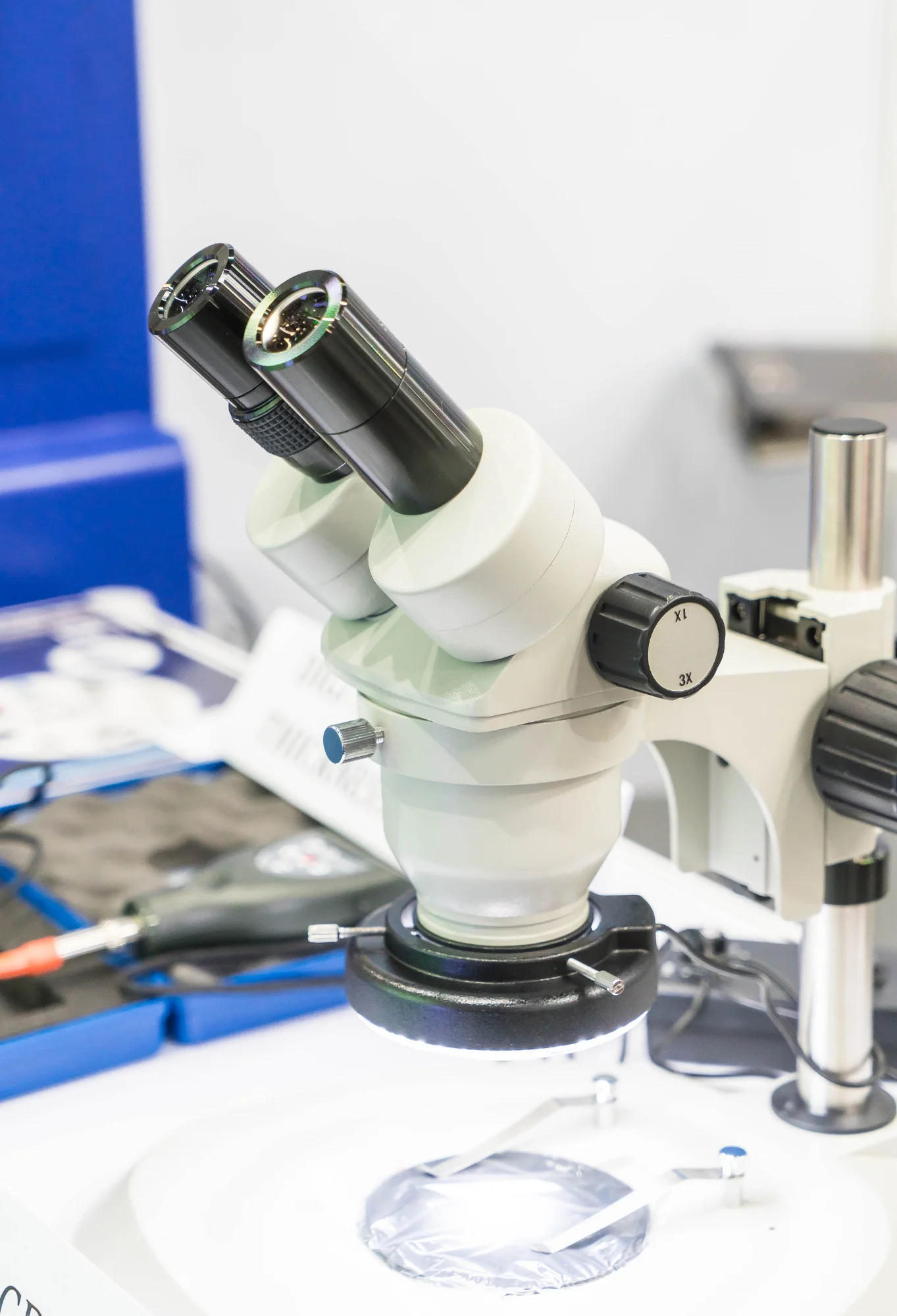
A breakthrough in volumetric imaging is transforming the landscape of biological research and analysis. In a groundbreaking study published in the journal Nature Methods, researchers from the University of California, San Francisco (UCSF) presented a novel epi-illumination single-plane illumination microscope (SPIM) system that combines high spatial-temporal resolution with subcellular precision and single-molecule sensitivity. This innovative system is set to revolutionize the way we observe and understand the complex dynamics of life at the microscopic level.
DOI: 10.1038/s41592-019-0401-3
The research team, led by Bo Huang, has developed an epi-illumination SPIM platform that achieves unparalleled imaging depth and quality using a single objective lens and an inverted fluorescence microscope interface. This method is streamlined and compatible with traditional biological sample holders, including multi-well plates, making it a versatile tool for cellular observation.
The study entitled “Epi-illumination SPIM for volumetric imaging with high spatial-temporal resolution” illustrates how the team’s design circumvents the limitations of conventional fluorescence microscopy by avoiding the need for additional reflection elements. This results in clearer, more detailed images that can contribute to a better understanding of various biological processes.
Bin Yang, Xingye Chen, Yina Wang, and others in the research group demonstrated the capabilities of the epi-illumination SPIM by conducting fast multicolor volumetric imaging, single-molecule localization microscopy, and parallel imaging of cellular responses across 16 different cell lines. These experiments illustrated the system’s potential for high-throughput analysis, a valuable asset for drug screening and fundamental biological research.
The UCSF-developed technology represents a significant leap forward in microscopy, particularly when analyzing living cells. The epi-illumination SPIM system allows researchers to observe cell behavior in three-dimensional space over time, with minimal disturbance to the specimens.
The development of the epi-illumination SPIM was published in Nature Methods under the following reference:
Yang, B., Chen, X., Wang, Y., Feng, S., Pessino, V., Stuurman, N., … & Huang, B. (2019). Epi-illumination SPIM for volumetric imaging with high spatial-temporal resolution. Nature Methods, 16(6), 501-504.
This research received funding from various NIH grants, suggesting the importance the National Institute of Health places on such technological advancements in biomedical science.
While discussing conflicts of interest, it is noted that a patent application has been filed for the reported microscope design.
The advantages of the epi-illumination SPIM system are manifold
1. Compatibility: The system fits seamlessly into existing microscopy workflows, as it is designed to interface with the sample holders already in use in biological labs around the world.
2. Versatility: The ability to perform multicolor imaging and single-molecule localization microscopy means that researchers can apply the system to a wide range of studies.
3. High Throughput: The design allows for parallel imaging, making it suitable for screening applications whereby numerous samples need to be analyzed quickly and efficiently.
4. Preservation of Sample Integrity: The system’s non-invasive nature means that it can image living cells over extended periods without damaging them, allowing for real-time observation of biological processes.
5. Superior Resolution: Epi-illumination SPIM’s spatial-temporal resolution enables researchers to observe the minutest details of cellular processes, down to single-molecule events.
For the scientific community, the implications of this new imaging technique are vast. It holds the promise of pushing the boundaries of cell biology, neuroscience, developmental biology, and a host of other fields where understanding the behavior of cells is crucial.
Reflecting on the impact of these findings, the scientific community refers to a series of references that underscore the importance of advanced imaging techniques in biological research, including works by Power RM & Huisken J, Huisken J et al., Tomer R et al., Wu Y et al., and more. The continued evolution of microscopy technology as demonstrated by this study not only showcases the incredible potential for new discoveries but also the ingenuity of scientists dedicated to expanding our ability to visualize life at the cellular level.
Keywords
1. Epi-illumination SPIM
2. Volumetric imaging technology
3. High-resolution cellular imaging
4. Single-molecule sensitivity microscopy
5. Advanced fluorescence microscopy techniques
References
1. Power RM & Huisken J (2017). A guide to light-sheet fluorescence microscopy for multiscale imaging. Nat. Methods, 14, 360–373.
2. Huisken J, Swoger J, Bene FD, Wittbrodt J & Stelzer EHK (2004). Optical sectioning deep inside live embryos by selective plane illumination microscopy. Science, 305, 1007–1009.
3. Tomer R, Khairy K, Amat F & Keller PJ (2012). Quantitative high-speed imaging of entire developing embryos with simultaneous multiview light-sheet microscopy. Nat. Methods, 9, 755.
4. Wu Y et al. (2011). Spatially isotropic four-dimensional imaging with dual-view plane illumination microscopy. Proc. Natl. Acad. Sci. U. S. A, 108, 17708–17713.
5. Holekamp TF, Turaga D & Holy TE (2008). Fast three-dimensional fluorescence imaging of activity in neural populations by objective-coupled planar illumination microscopy. Neuron, 57, 661–672.
Additional references supporting the creation and use of the epi-illumination SPIM system include discoveries and studies from a range of scientific investigations that detail the advancements in fluorescence microscopy and the novel application of imaging techniques in the life sciences.
In conclusion, the development of the epi-illumination SPIM system by Huang and colleagues is a major stride forward in microscopy. Its potential for significant contributions to the visualization of biological processes, understanding of disease mechanisms, and advancement of drug discovery is immense. As this technology becomes more widespread, we can expect a surge in groundbreaking biological insights, contributing to an ever-growing compendium of knowledge in the field.
