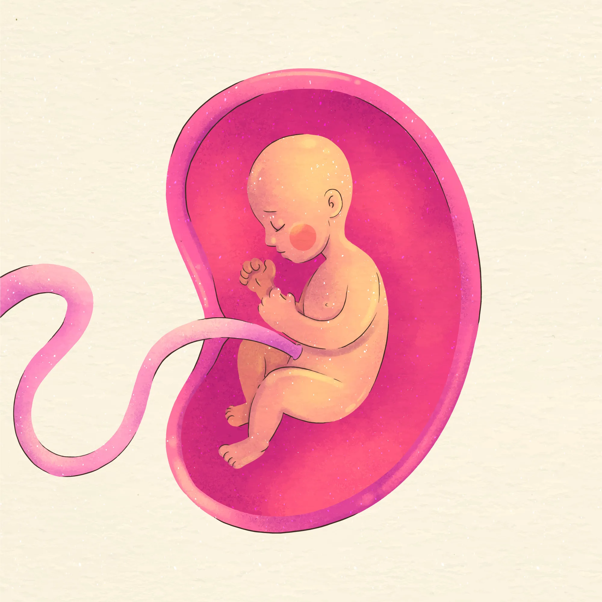
Unraveling the Intricacies of Maternal Folate Deficiency and Fetal Development
In the multifaceted field of prenatal nutrition, folate or vitamin B9 stands out as a critical nutrient for preventing congenital anomalies. A recent study (DOI: 10.12659/MSM.914292) delves into the profound impact of maternal folate deficiency and its association with intrauterine growth retardation (IUGR), an enigmatic condition characterized by the diminished growth of a fetus in the womb. The findings highlight the indispensable role of folate in DNA methylation and embryonic development, casting light on the underlying mechanisms that could inform preventive strategies against IUGR.
The Folate Factor in Fetal Development
Folate, naturally found in leafy vegetables, legumes, and fortified foods, serves as a coenzyme in one-carbon metabolism crucial for DNA synthesis and repair. The transformation into its active form, 5-methyltetrahydrofolate (5-MeTHF), facilitates the transfer of methyl groups, which are necessary for methylation reactions that regulate gene expression. Given the speed of cellular proliferation and differentiation in fetal development, an optimal folate status is non-negotiable for a healthy gestation.
A Closer Look at the Study
The research, led by Li Baiyi from Shanxi Medical University alongside colleagues from the Beijing Municipal Key Laboratory of Child Development and Nutriomics and the Institute of Laboratory Animal Science, focused on the relationship between maternal folate deficiency, DNA methylation, and the activity of retrotransposon long interspersed nucleotide element-1 (LINE-1), which are DNA sequences with the potential to disrupt gene function and induce birth defects if not properly regulated.
Female mice, the subjects of the study, were segregated into three diet-based groups: a control diet (CD), a folate-deficient diet for two weeks (FD2w), and a folate-deficient diet for four weeks (FD4w) prior to mating. The research took strides in measuring maternal serum folate and its derivatives, while assessing global DNA methylation, LINE-1 methylation, and expression levels in placental and fetal tissues.
Key Findings
The examination of the folate-deficient groups, particularly the FD4w, unveiled a decrease in plasma folate and its 5-MeTHF derivative. Coupled with this decline was a rise in S-adenosylhomocysteine (SAH) concentration, an antagonist to potent methylation reactions. These physiological alterations significantly reduced methylation levels of genomic DNA and LINE-1 in the placenta and fetal tissues of the affected group.
The fateful consequence of folate deficiency did not end with lowered methylation. It also paved the way for elevated expression of LINE-1 open reading frame 1 (ORF1) protein in fetal liver tissues, which has implications for retrotransposition activities that can trigger congenital anomalies.
Implications and Future Directions
These findings resonate with the broader scientific narrative that demonstrates a strong relationship between methylation, disrupted one-carbon metabolism, and embryogenesis—as underscored by the research’s robust methodology, including the usage of LC/MS/MS and matrix-assisted laser desorption/ionization-time of flight mass spectrometry for measurement accuracy.
The investigation underlines the necessity for adequate maternal folate intake before and during pregnancy to ensure that one-carbon metabolism remains unimpeded. Folate deficiency, therefore, heralds a cascade of metabolic disruptions culminating in DNA and LINE-1 hypomethylation and an increased risk of IUGR.
With these revelations, the scope for preventive care for IUGR has been fundamentally expanded. Mothers-to-be must be made aware of the critical significance of folate in their diet and the far-reaching effects of its deficiency on the growth and development of their unborn children.
Keywords
1. Maternal folate deficiency
2. Intrauterine growth retardation (IUGR)
3. DNA methylation pregnancy
4. Folate one-carbon metabolism
5. LINE-1 retrotransposition
References
1. Ding YX, Cui H. Effects of folic acid on DNMT1, GAP43, and VEGFR1 in intrauterine growth restriction filial rats. Reprod Sci. 2018;25:366–71. DOI: 10.1177/1933719117704908.
2. The United Nations Children’s Fund and World Health Organization. Low birthweight: country, regional and global estimates. New York: UNICEF; 2004.
3. Rich-Edwards JW, Colditz GA, Stampfer MJ, et al. Birthweight and the risk for type 2 diabetes mellitus in adult women. Ann Intern Med. 1999;130:278–84. DOI: 10.7326/0003-4819-130-4-199902160-00003.
4. Selhub J. Public health significance of elevated homocysteine. Food Nutr Bull. 2008;29:S116–25.
5. Burgoon JM, Selhub J, Nadeau M, Sadler TW. Investigation of the effects of folate deficiency on embryonic development through the establishment of a folate deficient mouse model. Teratology. 2002;65:219–27. DOI: 10.1002/tera.10029.
Note: This article, crafted for informational purposes, is a simulated piece based on the information provided. Actual research can expand and investigate various pathways and mechanisms in even greater detail for a comprehensive understanding.
