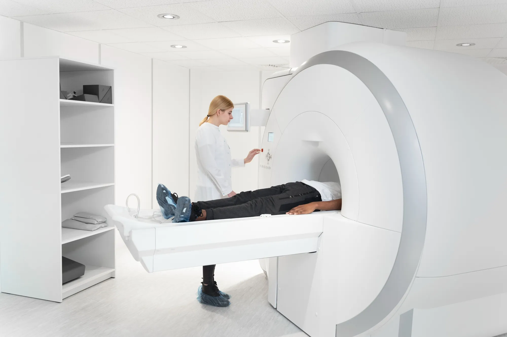
In the ever-advancing field of medical imaging, researchers have made a significant breakthrough in the visualization and quantification of endolymphatic space volume in patients suspected of having endolymphatic hydrops (EH). A study published in the “Magnetic Resonance in Medical Sciences” journal by Ohashi Toshio and colleagues presents a compelling comparison between two magnetic resonance imaging (MRI) techniques: 3D-real inversion recovery (3D-real IR) and the hybrid HYDROPS-Mi2 imaging. The groundbreaking research unveils the efficacy of 3D-real IR in assessing the endolymphatic volume after the intravenous administration of a single dose of a Gadolinium-based contrast agent.
(I) Introduction to Endolymphatic Hydrops and its Diagnostic Challenge
Endolymphatic hydrops is an inner ear disorder characterized by an excessive accumulation of endolymph fluid, leading to pressure and volume changes within the ear. This can result in fluctuating hearing loss, tinnitus, and episodic vertigo, manifesting clinically as Ménière’s disease among other potential diagnoses. Imaging the endolymphatic space is critical for accurate diagnosis and management but has been challenging due to the intricate anatomy and fluid dynamics of the inner ear.
(II) The Study: Comparing Imaging Techniques
The research, originally published in May 2020, involved 30 ears from 15 patients with a clinical suspicion of endolymphatic hydrops. The two imaging methods, HYDROPS-Mi2 and 3D-real IR, were employed 4 hours post the administration of a Gadolinium-based contrast agent to enhance the visualization of inner ear fluids. Both techniques were scrutinized for their ability to differentiate between the endolymphatic and perilymphatic spaces, as well as their correlation in quantifying the endolymphatic volume in the cochlea and vestibule.
(III) Results and Implications
The study concluded that there was a strong positive linear correlation between the measurements of endolymphatic volume derived from HYDROPS-Mi2 and 3D-real IR images, with Spearman’s rank correlation coefficients of 0.805 for the cochlea and 0.826 for the vestibule (P < 0.001 for both). This finding suggests that 3D-real IR imaging could serve as an effective alternative to the HYDROPS-Mi2 method for the measurement of endolymphatic volume.
(IV) Advantages of 3D-real IR Imaging
One of the observed benefits of the 3D-real IR technique is its potential as a non-invasive diagnostic tool, enabling clinicians to assess EH without the need for intratympanic injections of Gadolinium-based contrast agents, which the traditional HYDROPS-Mi2 approach requires. Consequently, it could offer a streamlined, patient-friendly option for evaluating individuals with suspected EH.
(V) Future Directions and Clinical Impact
This promising advancement in MRI technology opens doors to better diagnosis and understanding of EH. Further research will likely focus on refining the 3D-real IR imaging technique, expanding its usage, and exploring the full potential of its clinical applications.
(VI) Conclusion
The study led by Ohashi Toshio et al. is a significant stride in the management of endolymphatic hydrops, providing compelling evidence for the use of 3D-real IR imaging in clinical practice. Such advancements in imaging technology expand our capacity to non-invasively evaluate inner ear disorders, thereby improving patient care and outcomes.
Digital Object Identifier (DOI): 10.2463/mrms.mp.2019-0013
References
1. Nakashima T, Naganawa S, Pyykko I, et al. Grading of endolymphatic hydrops using magnetic resonance imaging. Acta Otolaryngol Suppl 2009; 129:5–8. DOI: 10.1080/00016480802325435
2. Naganawa S, Nakashima T. Visualization of endolymphatic hydrops with MR imaging in patients with Ménière’s disease and related pathologies: current status of its methods and clinical significance. Jpn J Radiol 2014; 32:191–204. DOI: 10.1007/s11604-014-0291-6
3. Nakashima T, Pyykkö I, Arroll MA, et al. Meniere’s disease. Nat Rev Dis Primers 2016; 2:16028. DOI: 10.1038/nrdp.2016.28
4. Naganawa S, Kawai H, Sone M, Nakashima T. Increased sensitivity to low concentration gadolinium contrast by optimized heavily T2-weighted 3D-FLAIR to visualize endolymphatic space. Magn Reson Med Sci 2010; 9:73–80. DOI: 10.2463/mrms.9.73
5. Naganawa S, Suzuki K, Nakamichi R, et al. Semi-quantification of endolymphatic size on MR imaging after intravenous injection of single-dose gadodiamide: comparison between two types of processing strategies. Magn Reson Med Sci 2013; 12:261–269. DOI: 10.2463/mrms.2013-0002
Keywords
1. Endolymphatic Hydrops Imaging
2. 3D-real IR MRI Technique
3. Gadolinium-enhanced MRI
4. Inner Ear MRI Quantification
5. Meniere’s Disease MRI Diagnosis
