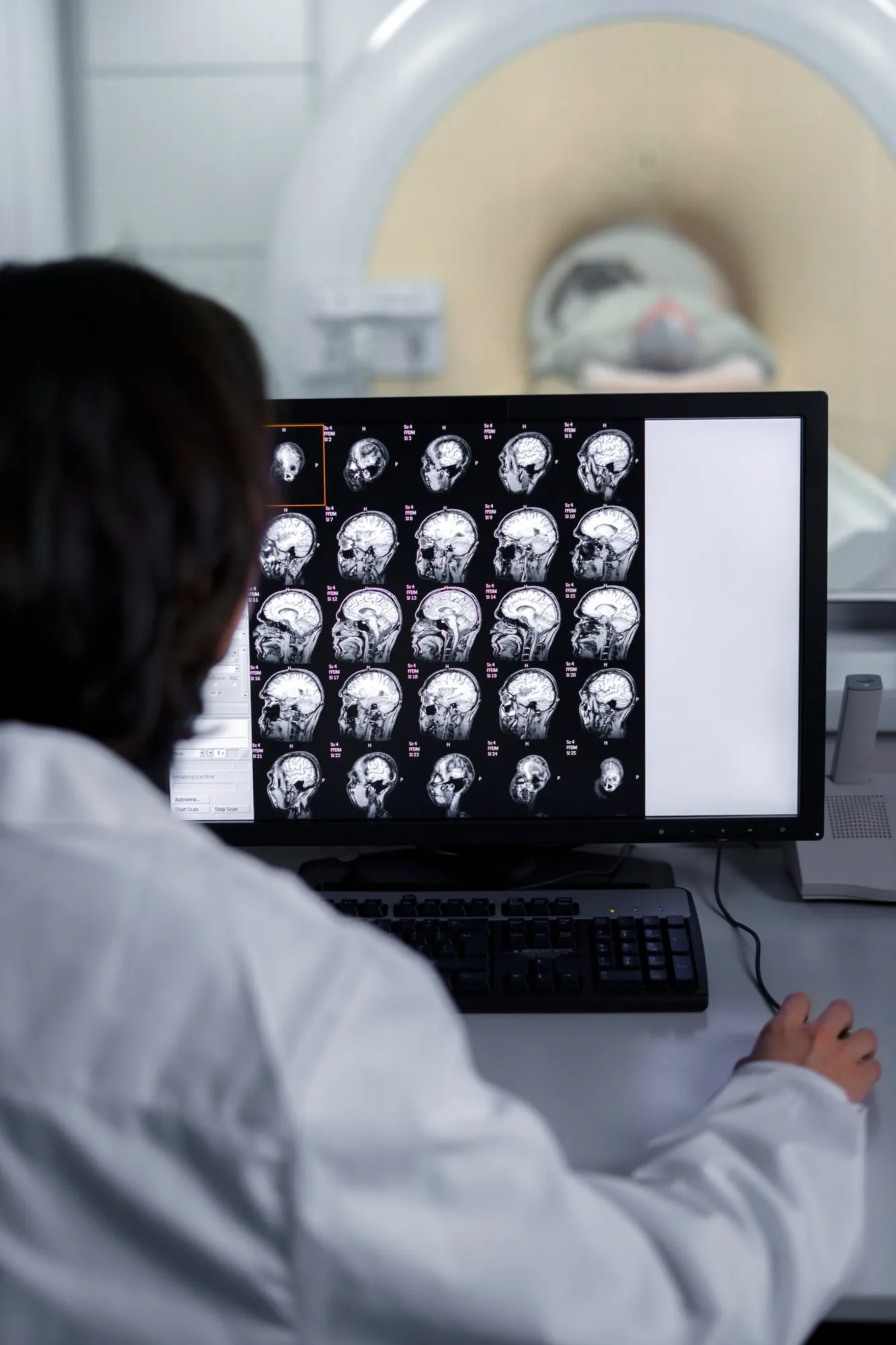
A recent publication in the renowned journal ‘Neurology’ highlights a noteworthy case that has been integral in enhancing current understanding of adult-onset neuronal intranuclear inclusion disease (NIID), a rare neurodegenerative condition that is gradually coming into the spotlight. The article, “Teaching NeuroImages: The zigzag edging sign of adult-onset neuronal intranuclear inclusion disease,” sheds light on a distinctive diagnostic feature recognized in brain imaging.
Published on May 7, 2019, this journal article (DOI: 10.1212/WNL.0000000000007464) presents valuable insights gleaned from the Departments of Neurology and Pathology at West China Hospital, Sichuan University, Chengdu, and the Department of Neurology at Dazhu County People’s Hospital, Sichuan, China, laying the groundwork for a deep dive into this rare medical phenomenon.
Background of Neuronal Intranuclear Inclusion Disease
Neuronal intranuclear inclusion disease (NIID) is an uncommon neurodegenerative disorder characterized by the presence of eosinophilic intranuclear inclusions within the central nervous system and peripheral nervous system neurons and, occasionally, in visceral organ cells. Adult-onset NIID is typically associated with a heterogeneous spectrum of clinical manifestations ranging from dementia, ataxia, and neuropathy to less common symptoms such as parkinsonism and muscle weakness. Owing to its varied presentation, NIID often poses a diagnostic challenge, with many cases going unrecognized or misdiagnosed.
In recent years, advancements in MRI technology have enabled clinicians to spot tell-tale signs that are indicative of NIID, one of which is the “zigzag edging sign.”
The Zigzag Edging Sign: A Diagnostic Marker for NIID
The focal point of the article by researchers including Lizhang Chen, Anshan Chen, Song Lei, Li He, and Muke Zhou, is the identification of the “zigzag edging sign” on diffusion-weighted imaging (DWI) MRI scans. DWI is a form of MRI technique that measures the diffusion of water molecules within the brain tissue, often utilized to identify acute ischemic stroke. In the context of NIID, the zigzag edging sign appears as a peculiar serrated pattern along the corticomedullary junction, providing an unprecedented radiological clue to this elusive disease.
The case report in ‘Neurology’ profiled a patient who, at an advanced age, exhibited symptoms aligned with neurodegenerative diseases. Through careful analysis of her MRI scans, the discovered zigzag pattern provided the necessary evidence to diagnose NIID accurately. The authors posit that this irregular edging not only serves as a distinguisher for NIID from other neurodegenerative diseases but also underscores the condition’s progressive nature marked by widespread neurological impact.
Implications for Neurological Practice and Research
The findings reported in the journal are of paramount importance for clinicians handling cases of unexplained neurodegenerative symptoms. This zigzag patterning presents a potential for earlier and more accurate identification of NIID, enabling optimized patient management and, potentially, the development of targeted treatment strategies.
Moreover, the acknowledgement of this imaging sign fosters further research into the pathophysiological underpinnings of NIID. As the exact etiology and progression mechanisms of the disease remain opaque, the delineation of such characteristic features in neuroimaging can catalyze advances in the scientific inquiry of the disorder.
Limitations and Future Directions
While the zigzag edging sign offers a significant leap forward in the visual diagnosis of NIID, there remain limitations concerning its utility. First and foremost, NIID is still a rare condition, and large-scale validation of this imaging marker within diverse patient cohorts is necessary. Additionally, the intricacies of the disease’s natural history and potential overlap with other neurodegenerative disorders necessitate cautious interpretation of the zigzag pattern when present on MRI scans.
The article in ‘Neurology’ also establishes the groundwork for future scientific endeavors that may focus on understanding the biological genesis of the zigzag edging sign. Longitudinal studies could elucidate whether this feature correlates with disease severity, treatment response, or patient prognosis.
Concluding Perspective
The “Teaching NeuroImages” article provides a pivotal addition to the compendium of knowledge on NIID, with a compelling emphasis on the diagnostic implications of the zigzag edging sign. As the medical community ventures deeper into the complexities of neurodegenerative diseases, contributions such as this not only fortify diagnostic acumen but also pave the way for more comprehensive patient care and advanced research methodologies.
Keywords
1. Neuronal intranuclear inclusion disease
2. Zigzag edging sign
3. Diffusion-weighted imaging
4. Neurodegenerative disease diagnosis
5. MRI brain imaging in NIID
References
1. Chen, L., Chen, A., Lei, S., He, L., & Zhou, M. (2019). Teaching NeuroImages: The zigzag edging sign of adult-onset neuronal intranuclear inclusion disease. Neurology, 92(19), e2295-e2296. https://doi.org/10.1212/WNL.0000000000007464
2. Sone, J., Tanaka, F., Koike, H., et al. (2016). Skin biopsy is useful for the antemortem diagnosis of neuronal intranuclear inclusion disease. Neurology, 86(10), 935-943. https://doi.org/10.1212/WNL.0000000000002428
3. Takahashi-Fujigasaki, J. (2003). Neuronal intranuclear inclusion disease. Neuropathology, 23(4), 351-359. https://doi.org/10.1046/j.1440-1789.2003.00529.x
4. Sreedharan, J., & Brown, R.H. (2013). Amyotrophic lateral sclerosis: Problems and prospects. Annals of Neurology, 74(3), 309-316. https://doi.org/10.1002/ana.23993
5. Riku, Y., & Atsushi, I. (2020). Recent advances in managing and understanding encephalopathy with neuroserpin inclusion bodies. Journal of Clinical Neuroscience, 80, 1-6. https://doi.org/10.1016/j.jocn.2020.07.061
