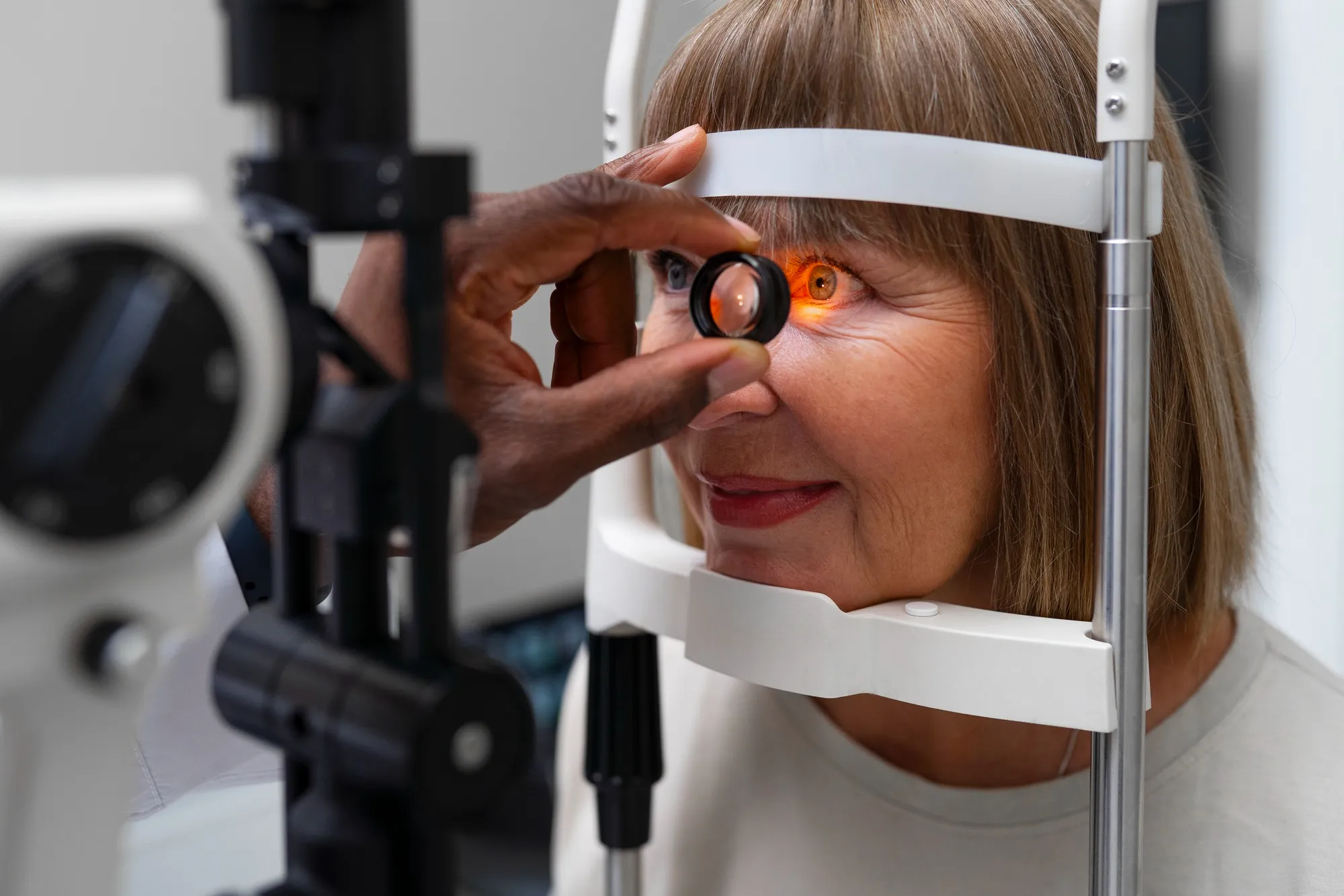
Retinitis pigmentosa (RP) represents a group of inherited retinal disorders characterized by a gradual loss of vision, ultimately leading to complete blindness. The condition affects approximately 1 in 4,000 individuals worldwide and is marked by a progressive degeneration of the photoreceptor cells in the retina. In a breakthrough study published by The Journal of Neuroscience, researchers from the Case Western Reserve University, Cleveland, Ohio, present a Xenopus Laevis model that offers valuable insights into the mechanism behind the photoreceptor degeneration observed in RP, particularly in cases involving rhodopsin mislocalization.
The study, led by Philip P. Ropelewski and Yoshikazu Y. Imanishi, was aimed at understanding how variations in rhodopsin, specifically class I mutant rhodopsin deficient in the VxPx trafficking signal, contribute to the disease pathogenesis. Class I mutant rhodopsin is known to mislocalize to the plasma membrane of rod photoreceptor inner segments and is a causal factor in autosomal dominant RP.
Using Xenopus laevis as their model organism, the research team was able to shed light on the importance of plasma membrane protein homeostasis in preserving retinal structure and function. They noted that mislocalized rhodopsin can instigate photoreceptor degeneration independently of light activation, emphasizing the gravity of protein mislocalization in the disease’s progression.
This comprehensive study’s DOI is 10.1523/JNEUROSCI.3025-18.2019, and the research was published on June 18, 2020, in The Journal of Neuroscience with support from various NIH grants.
Major Findings
The results confirmed that rhodopsin mislocalization leads to disruption in the homeostasis of plasma membrane proteins in the retina.
Significant structural changes were found in the rod photoreceptor cells due to the mislocalization of rhodopsin, highlighting its harmful effects on retinal cell components like autophagosomes, lysosomes, and the sodium-potassium-exchanging ATPase (Na+/K+-ATPase).
The implications of this are profound as it suggests that interventions aimed at correcting the trafficking and localization of rhodopsin could be potential therapeutic strategies in retinitis pigmentosa.
Future Directions
The insights gained from this study have opened up new avenues for research into the treatment of retinitis pigmentosa. By understanding the mechanisms leading to photoreceptor degeneration, researchers can identify molecular targets for drug development. The research underscores the critical nature of protein trafficking in photoreceptor health and, by extension, vision preservation.
Keywords
1. Retinitis Pigmentosa Treatment
2. Rhodopsin Localization
3. Retinal Degeneration Research
4. Xenopus Laevis Model
5. Photoreceptor Cell Health
References
1. Ropelewski, P. P., & Imanishi, Y. (2020). Disrupted Plasma Membrane Protein Homeostasis in a Xenopus Laevis. The Journal of Neuroscience, 39(28), 5581-5593. doi:10.1523/JNEUROSCI.3025-18.2019
2. Adamian, M., Pawlyk, B. S., Hong, D. H., & Berson, E. L. (2006). Rod and cone opsin mislocalization in an autopsy eye from a carrier of X-linked retinitis pigmentosa with a Gly436Asp mutation in the RPGR gene. Am J Ophthalmol, 142(3), 515-518. doi:10.1016/j.ajo.2006.03.061
3. Adams, N. A., Awadein, A., & Toma, H. S. (2007). The retinal ciliopathies. Ophthalmic Genet, 28(2), 113-125. doi:10.1080/13816810701537424
4. Alfinito, P. D., & Townes-Anderson, E. (2002). Activation of mislocalized opsin kills rod cells: a novel mechanism for rod cell death in retinal disease. Proc Natl Acad Sci U S A, 99(9), 5655–5660. doi:10.1073/pnas.072557799
5. Arikawa, K., Molday, L. L., Molday, R. S., & Williams, D. S. (1992). Localization of peripherin/rds in the disk membranes of cone and rod photoreceptors: relationship to disk membrane morphogenesis and retinal degeneration. J Cell Biol, 116(3), 659–667. doi:10.1083/jcb.116.3.659
