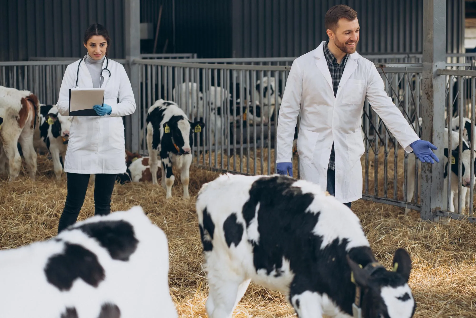
In veterinary medicine, the phenomenon of osteochondrosis (OC) in animals, particularly in swine, denotes a prevalent clinical challenge with substantial implications for animal health and the agricultural industry. A groundbreaking study published in ‘Veterinary Pathology’ provides in-depth insights into the initiation of osteochondrosis in the distal femoral physis of pigs, emphasizing its vascular origins and the pathogenesis akin to articular OC. Here we disseminate the study’s core findings, methodological details, and potential implications for veterinary diagnostics and animal breeding.
Conducted by a research team led by Kristin K. Olstad and colleagues at the Norwegian University of Life Sciences and Norsvin Delta, Hamar, Norway, the study aimed to elucidate the histological and computed tomographic (CT) characteristics of changes in the distal femoral physis of pigs, determining their correlation with OC lesions and their pathogenesis (DOI: 10.1177/0300985819843685). This article meticulously examines the research, accompanied by implications and future directives in the domain of veterinary OC research. The findings of this study not only contribute to a better understanding of OC in pigs but may also hold implications for OC in larger animal models and human medicine.
Introduction to Osteochondrosis in Pigs
Osteochondrosis is a developmental bone and cartilage disorder that affects the joint cartilage and the growth plate of animals, with a significant impact on pigs. This pathology can lead to pain, impaired movement, and potential economic loss due to reduced animal welfare and productivity. The disorder has been linked to issues in bone growth, specifically to disruptions in blood flow resulting in the necrosis of cartilage—a vital material responsible for the smooth function and structure of joints.
Study Methodology
The research material included 19 male Landrace pigs specifically bred for a predisposition to OC. Pigs were dutifully euthanized and CT-scanned at two-week intervals from 82 to 180 days of age to chronologically document the development of their femoral physes. From this group, 10 pigs provided material for subsequent histological validation to affirm the CT scan findings.
Key Findings
Through CT imagery, the researchers identified 31 lesions, which morphological examination later confirmed as compatible with OC. The lesions were primarily ascribed to the vascular disruption of end arteries—the microvascular structures supplying blood to the epiphysis, coursing towards the metaphysis. Notably, the study unveiled that the origin of lesions was in alignment with the expected failure point where blood vessels meld into the bone.
It was reported that vascular failure was associated with an unusual delay in ossification and the retention of viable hypertrophic chondrocytes, as opposed to cartilage necrosis, which often hallmarks OC lesions in proximal animals and even humans. Interestingly, the study did not observe any angular limb deformity in any of the pigs.
Discussion and Implications
The research by Olstad et al. portrayed osteochondrosis as a condition initiated by a vascular failure identical to what is observed in articular OC. If the hereditary predisposition is perceived to be constant across these locations, it may necessitate a revision of preventative breeding practices to mitigate the prevalence of OC.
The potential for some lesions to self-resolve due to small size and evidence of reparative ossification observed in CT scans indicates that early detection and monitoring might be pivotal in managing OC effectively. Moreover, the study indicates that the lesions’ impact on limb deformity and broader implications for the pig’s growth and development necessitate further research in older pigs.
References
1. Olstad, K. et al. (2019). Osteochondrosis in the Distal Femoral Physis of Pigs Starts With Vascular Failure. Veterinary Pathology, 56(5), 732-742. DOI: 10.1177/0300985819843685.
2. Ytrehus, B., Carlson, C. S., & Ekman, S. (2007). Etiology and pathogenesis of osteochondrosis. Veterinary Pathology, 44(4), 429-448.
3. Grøndalen, T. (1979). Osteochondrosis and arthrosis in pigs. Acta Radiologica Supplementum, 358, 299-305.
4. McIlwraith, C. W. (1988). Osteochondrosis. Veterinary Clinics of North America: Equine Practice, 4(2), 279-298.
5. Dik, K. J., Enzerink, E., & van Weeren, P. R. (1999). Radiographic development of osteochondrosis dissecans in the horse. Equine Veterinary Journal, 31(5), 369-374.
Keywords
1. Swine Osteochondrosis Research
2. Veterinary Bone Pathology
3. Pig Growth Plate Disorders
4. Farm Animal Joint Health
5. Animal Cartilage Disease Imaging
In conclusion, the study establishes a vital connection between the vascular failure and osteochondrosis in pigs, highlighting the inherent nature of this condition, with an emphasis on early detection and potential management strategies. As the veterinary and livestock industry continue to expand, the significance of robust and informed approaches to animal welfare and productivity aligned with such research insights cannot be overstated. This study not only reaffirms the role of specialized imaging in diagnosing OC but also sets a directive for continuous research in older swine populations, aiming to delineate the link between early physiological changes and wider implications for animal health.
