The complexity of epilepsy has challenged the medical community for decades. Identifying precise brain regions responsible for epileptic seizures—a concept known as the epileptogenic zone (EZ)—remains a pivotal task for neurosurgeons. The traditional method of localizing these zones through ictal electroencephalography (EEG) during seizures has its limitations due to the unpredictable nature of seizure events. However, a breakthrough in the field emerges as researchers turn to interictal intracranial EEG recordings to potentially pinpoint EZs. In an insightful study recently published in “Neurologia medico-chirurgica,” Nagata Keisuke et al. explore the promise of excitatory and inhibitory neuronal activity as biomarkers for epileptogenicity.
Interictal EEG: A New Horizon in Epilepsy Research
The study explores the basis of crucial electrophysical patterns that surface between seizures—referred to as interictal periods—which could serve as indicators of the epileptogenic zone. Identifying and interpreting these patterns could transform the current approach to epilepsy surgery.
A significant element of this research involved high-frequency oscillations (HFOs), initially considered a robust biomarker for abnormal excitatory activity in the ictogenic region. Despite the initial excitement, HFO-guided resections did not yield an improved seizure prognosis as hoped. The unexpected outcome prompted researchers to probe deeper into the mechanics of excitatory and inhibitory activity within the brain.
The team from the University of Tokyo, alongside researchers from Jichi Medical University, turned their focus to low-gamma oscillations (30-80 Hz), which seem to indicate hypersynchronization of inhibitory interneurons. They observed that assessing the regularity of these oscillations could facilitate the localization of the EZ with greater precision.
The Advent of Corticocortical Evoked Potentials (CCEP)
Aside from resting-state EEG assessments, an innovative modality named corticocortical evoked potentials (CCEP) made a considerable impact on evaluating epileptogenic territories. CCEP involves single-pulse electrical stimulation to elicit evoked potentials, which in turn, reflect functional connectivity within the brain.
When responses were recorded in regions remote from the stimulus site, researchers could glean evidence of intricate connectivity, notably an increase within the ictogenic region. This heightened internal connectivity and elevated inhibitory input from non-involved areas into the ictogenic zone provided a more nuanced understanding of the epileptic network.
In contrast, powerful responses noted near the stimulation site were indicative of local neuronal excitability, showcasing itself through an increased N1 amplitude and overpowering HFO. This knowledge opens up a fascinating avenue for recognizing the excitatory and inhibitory balance, which is critical for developing a surgical strategy for the treatment of epilepsy.
Further Research and Implications
Nagata Keisuke et al. call for further research to ascertain whether these novel electrophysiological approaches could serve, either individually or synergistically, as reliable biomarkers of epileptogenicity. The potential of these breakthroughs to improve seizure prognosis is substantial.
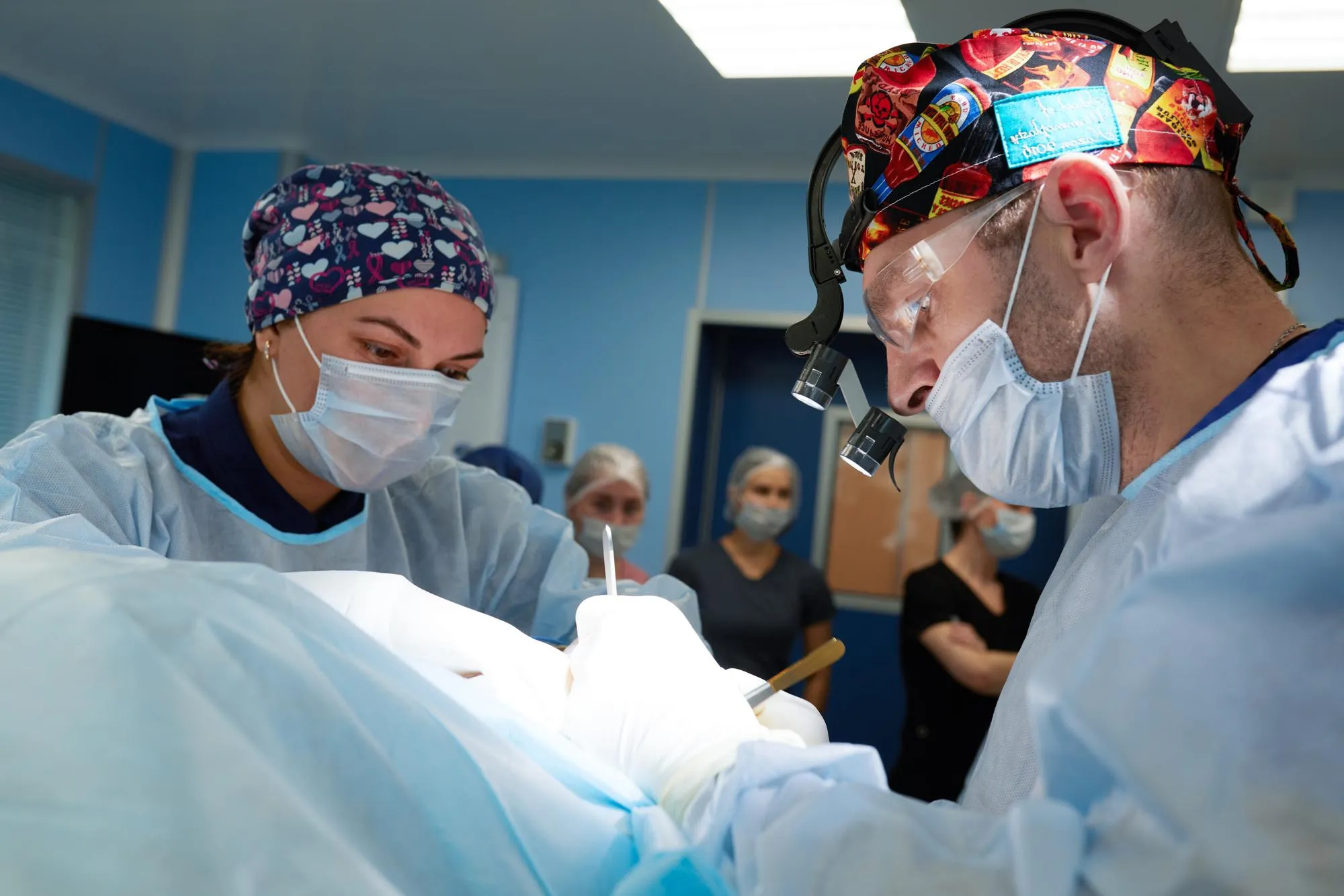
As the study transitions from theory to potential clinical application, the findings underscore an important shift in the landscape of epileptic surgery. The identification of non-invasive biomarkers through EEG would equip surgeons with the information needed to execute highly targeted resections. This could diminish the risks and raise the success rate of surgeries intended to eliminate or significantly reduce the occurrence of epileptic seizures.
It is imperative that follow-up studies continue to validate these findings and streamline the methods into practical techniques that could be ubiquitously applied in clinics and hospitals worldwide. Only through rigorous investigation and the refinement of these methods can a new standard of care emerge for individuals with epilepsy.
DOI:
The DOI for this study is 10.2176/jns-nmc.2023-0207.
References
1. Nagata, K., Kunii, N., Shimada, S., & Saito, N. “Utilizing Excitatory and Inhibitory Activity Derived from Interictal Intracranial Electroencephalography as Potential Biomarkers for Epileptogenicity.” Neurol Med Chir (Tokyo). 2024 Jan 15; 10.2176/jns-nmc.2023-0207.
2. Engel, J. “What’s in a name? The terminological confusion between epileptogenic/effectual/functional/lesional and symptomatic/cryptogenic/idiopathic/generalized seizures and their respective classifications.” Epilepsia, 2006; 47(10):1629-1634. DOI:10.1111/j.1528-1167.2006.00722.x.
3. Zijlmans, M., Jacobs, J., Zelmann, R., Dubeau, F., & Gotman, J. “High-frequency oscillations mirror disease activity in patients with epilepsy.” Neurology, 2009; 72(11):979-986. DOI:10.1212/01.wnl.0000343008.58319.aa.
4. Heers, M., Rampp, S., Stefan, H., Urbach, H., Elger, C. E., & von Lehe, M. “Merging of high frequency oscillation during presurgical epilepsy monitoring.” J Neurol Neurosurg Psychiatry, 2010; 81(5):529-534. DOI:10.1136/jnnp.2009.183517.
5. Matsumoto, A., Brinkmann, B. H., Stead, S. M., Matsumoto, J., Kucewicz, M. T., Marsh, W. R., Meyer, F. B., Worrell, G. A. “Pathological and physiological high-frequency oscillations in focal human epilepsy.” J Neurosurg, 2013; 119(5):1236-1246. DOI:10.3171/2013.7.JNS12214.
Keywords
1. Epileptogenic Zone EEG Biomarkers
2. Intracranial EEG Epilepsy Surgery
3. Excitatory Inhibitory Neuronal Activity
4. High-Frequency Oscillation Epilepsy
5. Corticocortical Evoked Potentials Epilepsy
