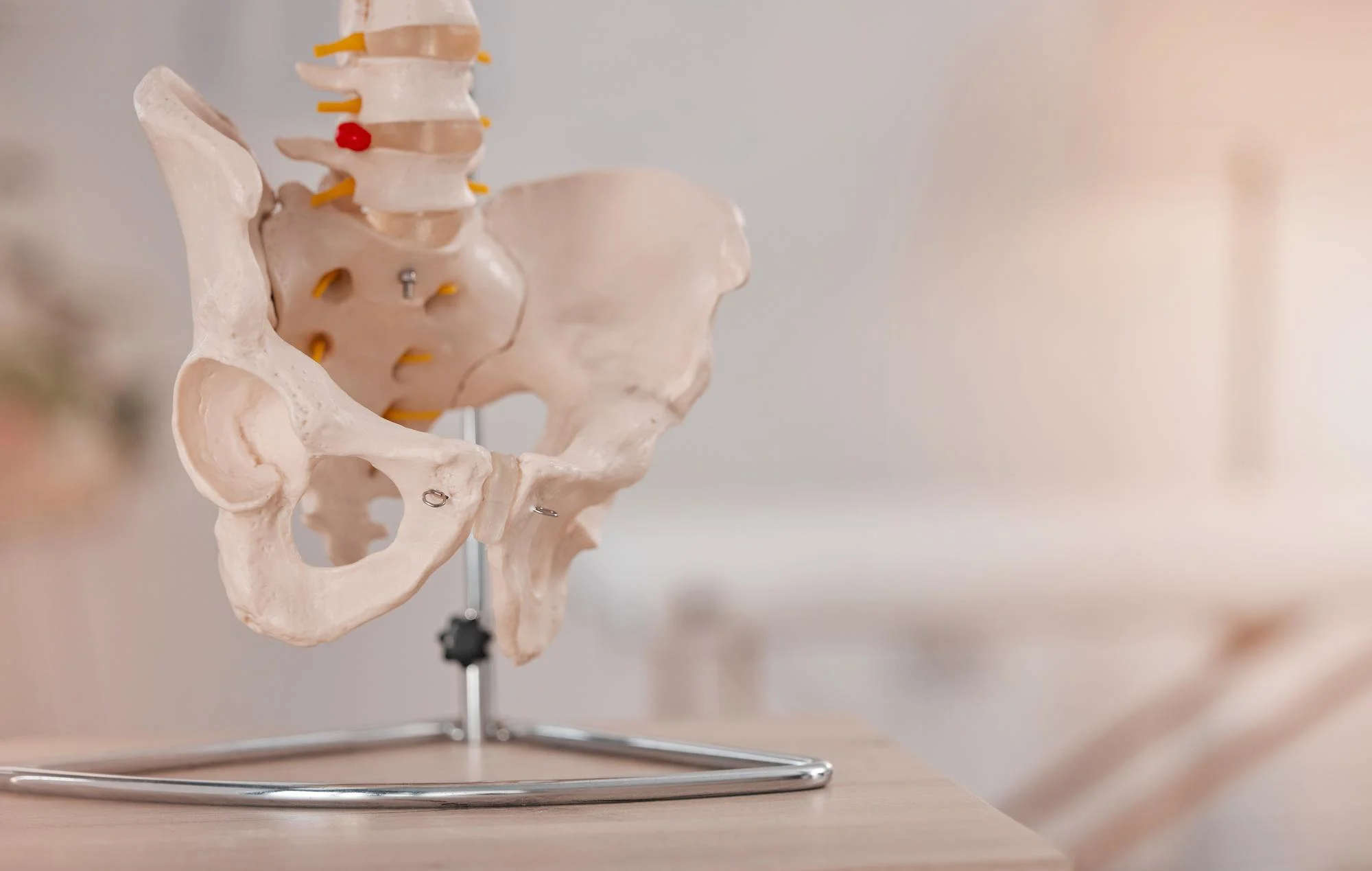
The complex and nuanced relationship between our spinal structure and overall physiology is a subject of enduring research, scrutiny, and discussion within the medical community, particularly in the sophisticated realms of neurosurgery and orthopedics. A study published on January 13, 2024, in the esteemed journal ‘World Neurosurgery’, probes the associations of transitional lumbosacral vertebrae (TLSV) – specifically sacralization and lumbarization – with spinopelvic parameters and lumbar lordosis (LL). Leading the investigation, Dr. Hamza H Karabag from Harran Üniversity in Şanlıurfa, Turkey, alongside Dr. Ahmet Celal Iplikcioglu of BHT Clinic Istanbul Tema Hastanesi, puts forth groundbreaking insights into this anatomical conundrum, invoking significant implications for both clinical assessments and potential interventions in spinopelvic health.
DOI: 10.1016/j.wneu.2024.01.056
The Anatomy of TLSV: Interpreting the Spinopelvic Language
Transitional lumbosacral vertebrae (TLSV) occur when there is a variation in the formation of the lumbosacral joint where the lumbar spine transitions into the sacral spine. These variants come in two primary forms: sacralization, where a lumbar vertebra is fused to the sacrum, and lumbarization, which is the non-fusion of the first sacral segment to the rest of the sacrum, essentially creating an additional movable lumbar vertebra.
Exploring the Parameters: The Upper and Lower Discourse
The World Neurosurgery study provides an analytical lens to view spinopelvic parameters through the scope of upper and lower sacral endplates in the context of TLSV. What is worth noting is that affected patients showcase two sets of values for these parameters and LL, warranting a tailored approach to their evaluation. The investigation reticulated data from 1420 asymptomatic patients, 108 of whom possessed TLSV, measuring their spinopelvic parameters and LL via abdominal computed tomography (CT).
Sacralization vs. Lumbarization: A Comparative Stance
Upon close examination, distinct variations became apparent. When referring to upper endplate measurements, individuals presented with lower spinopelvic parameters and LL values than the normative group. Conversely, lower endplate measurements revealed heightened values. Intriguingly enough, despite their anatomical differences, the lumbarization and sacralization groups manifested similar parameters when compared to each other.
Correlation and Consistency: The Numeric Dance
The rigorous analysis extended to probe the correlation between upper and lower endplate parameters. The results exhibited a robust relationship between them. A relatively constant difference was discerned between the upper and lower pelvic incidence (PI) and LL values, recorded at 27° and 14°, respectively. This constant sheds light on the considerable uniformity in the spatial placement of a sacralized L5 and a lumbarized S1 within the pelvic structure. It asserts that either parameter can be effectively harnessed for radiological assessments.
Implications and Future Directions: Beyond Asymptomatic Patients
Despite the study’s insights, Drs. Karabag and Iplikcioglu emphasize the necessity of extending research to individuals with TLSV-related symptoms. The analysis has predominantly hovered around asymptomatic subjects, potentially leaving gaps regarding how these spinopelvic dynamics manifest clinically and affect overall sagittal balance and quality of life.
The novel findings underscore the importance of recognizing these anatomical variants and their implications for patient care. Medical professionals may need to adopt modified evaluation strategies for patients presenting with TLSV to ensure proper diagnosis, tailor treatment plans, and steer rehabilitative endeavours for optimized outcomes.
Advancing Clinical Perspectives: A Reflective Afterword
The exploration of sacralization and lumbarization within the framework of spinopelvic parameters stands as a testament to the continuous advancements in spinal health research. The harmonization of vertebral form and function reverberates through the corridors of global neurosurgical and orthopedic practices, insisting on a refined comprehension of our anatomical idiosyncrasies.
Copyright Information
This comprehensive work, documented under the identifier S1878-8750(24)00068-8 and DOI 10.1016/j.wneu.2024.01.056, is safeguarded by copyright © 2024 Elsevier Inc. All rights held with utmost responsibility and integrity.
Keywords
1. Transitional lumbosacral vertebra
2. Spinopelvic parameters
3. Lumbar lordosis
4. Sacralization and lumbarization
5. Spinopelvic balance
References
1. Karabag, H. H., & Iplikcioglu, A. C. (2024). Analysis of Spinopelvic Parameters and Lumbar Lordosis in Patients with Transitional Lumbosacral Vertebrae, with Special Reference to Sacralization and Lumbarization. World Neurosurgery. https://doi.org/10.1016/j.wneu.2024.01.056
2. Labelle, H., Roussouly, P., Berthonnaud, E., Transfeldt, E. E., O’Brien, M., Chopin, D., … & Hresko, M. T. (2005). Spondylolisthesis, pelvic incidence, and spinopelvic balance: a correlation study. Spine, 30(18), 2049-2054.
3. Vialle, R., Levassor, N., Rillardon, L., Templier, A., Skalli, W., & Guigui, P. (2005). Radiographic analysis of the sagittal alignment and balance of the spine in asymptomatic subjects. The Journal of bone and joint surgery. American volume, 87(2), 260-267.
4. Roussouly, P., Gollogly, S., Noseda, O., Berthonnaud, E., & Dimnet, J. (2006). The vertical projection of the sum of the ground reactive forces of a standing patient is not the same as the gravity line: A radiographic study of the sagittal alignment of 153 asymptomatic volunteers. Spine, 31(11), E320-E325.
5. Bao, H., Vargas, L., Su, P., Vanden Berghe, M., & Lafage, V. (2019). Review of Spinal Parameters and Modular Implants for Correcting Adult Spinal Deformity. Clinical Spine Surgery, 32(8), 324-330.
